The short answer: a protein-binding result, not a proven treatment
IGF-1 DES, written des(1–3)IGF-I in the research literature, is a shortened form of insulin-like growth factor 1 (IGF-I). Investigators have examined whether deleting its first three amino acids changes how it interacts with insulin-like growth factor-binding proteins (IGFBPs). In several experimental systems the analogue escaped binding by the specific proteins tested while retaining access to the type 1 IGF receptor (IGF1R). Those are biochemical and cell-model findings, not evidence that a person will grow muscle, recover an injury or avoid harm. [1] Ross et al. [2] Remacle-Bonnet et al..
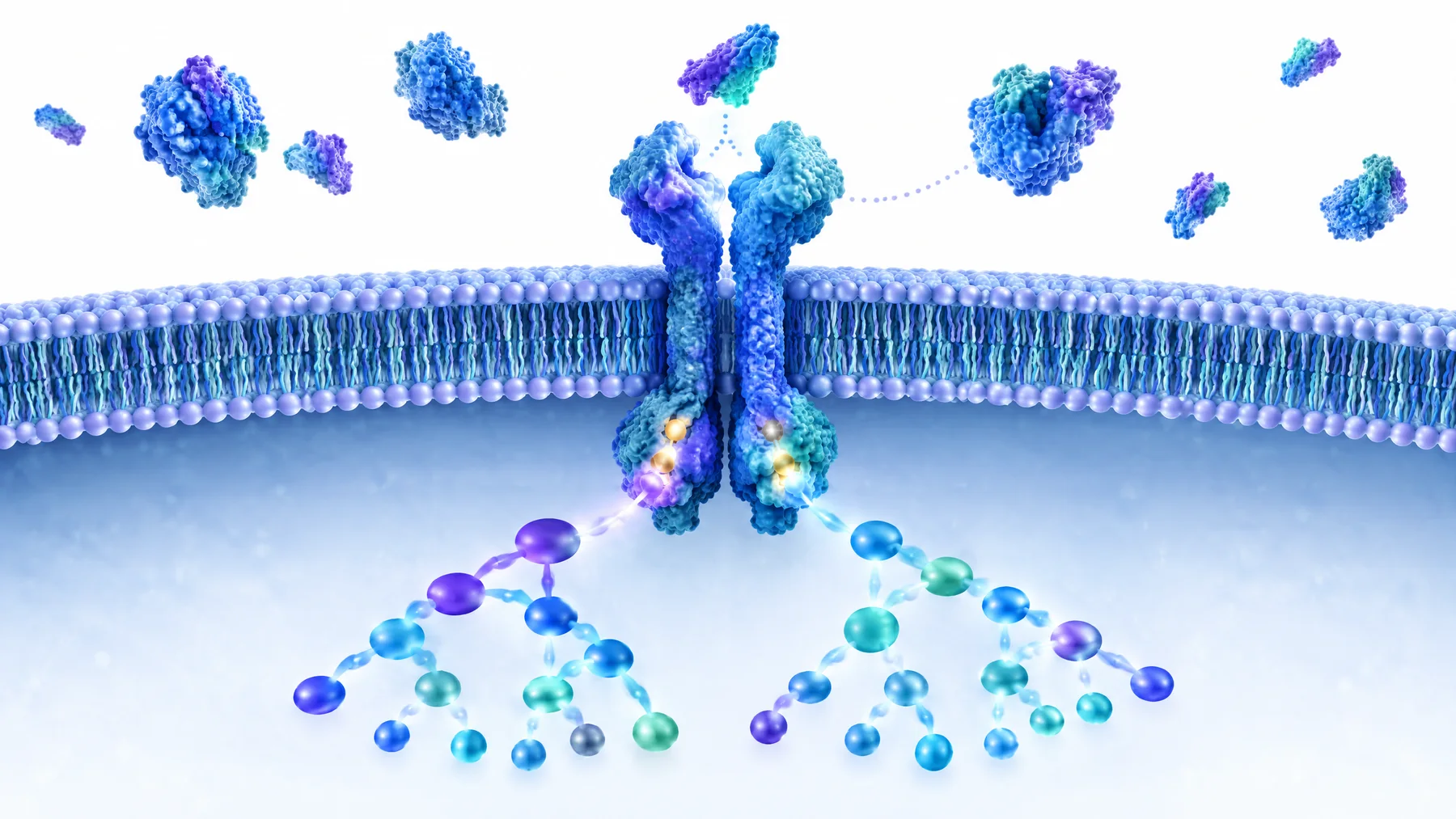
The image shows an illustrative receptor and signalling network, not a picture of a measured effect in a human subject. Under it, our labelled mechanism panel makes the boundary between observed and inferred steps explicit.
What does the ‘DES’ part mean?
Native human IGF-I has 70 amino acids. The DES variant lacks residues 1–3 at its amino-terminal end, leaving 67 amino acids; those missing residues are Gly–Pro–Glu. The N-terminal change matters because parts of this region contribute to binding-protein interactions. IGFBPs regulate how growth factors move, persist and become available to receptors in different tissues. A finding of weaker binding in one assay does not mean the protein has no binding partners anywhere in a living body. [3] Structural review.
The terminology can be confusing: IGF-I is the molecule, IGF1R is a cell-surface receptor, and an IGFBP is a regulatory binding protein. They are distinct actors. ‘DES’ is not a special kind of insulin syringe or a clinical designation; it is a shorthand for the molecular deletion.
The original binding-protein experiment
In a 1989 biochemical study, Ross and colleagues tested an IGF-binding protein purified from bovine-kidney cells using chick embryo fibroblasts and other experimental systems. The protein reduced several IGF-I and IGF-II activities but did not similarly reduce the biological activity of des(1–3)IGF-I in their tested setting. The researchers connected the analogue's greater observed potency there to its reduced interaction with the tested binding proteins. [1] Primary paper.
This does not support a universal ‘ten times stronger’ label, a predicted duration of action, a clinical dose, or a claim that all IGFBPs always suppress IGF action. Even that paper points to previously reported circumstances in which a binding protein enhanced rather than inhibited a response. When receptor environment, protein type, tissue or endpoint changes, the observed relationship can change too.
A human cell line is not a human trial
Another study used HT29-D4 human colon-carcinoma cells. Binding proteins secreted by these cultured cells did not bind des(1–3)IGF-I in the authors' assays, while cell-surface IGF1R bound native IGF-I and DES similarly. The DES variant was used as a laboratory tool to examine an IGF signalling loop. Some differentiation-related changes occurred in that cell model. [2] Primary paper.
The cells were human-derived, but that does not make the experiment a study of DES treatment in patients. It cannot establish tissue targeting, athletic performance, useful muscle gain, a cancer treatment, general safety or an adverse-event rate. The cell-line identity matters precisely because an apparently ‘human’ experimental material can still be far removed from a clinical outcome.
What happens at IGF1R? Follow the two main branches
IGF1R is a receptor tyrosine kinase. When a suitable ligand binds, the receptor's intracellular portions undergo phosphorylation and recruit adaptor molecules such as IRS proteins and Shc. The IRS–PI3K–AKT–mTOR axis participates in survival, metabolism and protein-synthesis-related signalling; the Shc–RAS–MAPK axis contributes to growth and differentiation-related signalling. These are common IGF1R biology pathways, not DES-specific clinical endpoints. [4] IGF1R signalling review.
The proposed relationship is: in some experimental environments, a reduced tendency to be captured by certain IGFBPs may leave more DES available to interact with IGF1R. Whether, where and for how long this occurs in a person's tissues is a separate pharmacokinetic question. No verified evidence here supports an arrow from the molecule directly to ‘localised muscle growth’ or ‘safe recovery’ in humans.
Why the same diagram can mean different things in different systems
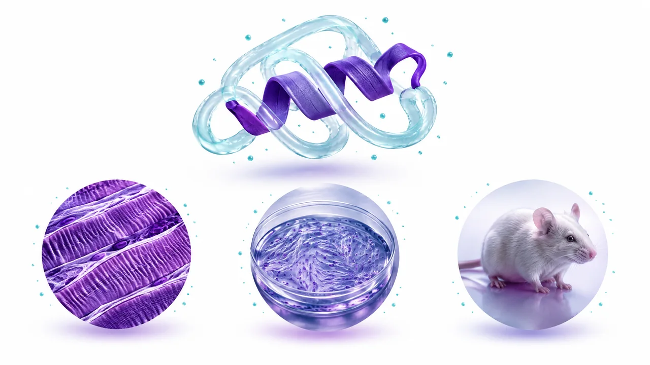
Think of an IGFBP as a molecular interaction partner, not a single universal lock. There are several binding proteins, and their effects depend on concentration, location, time and the assay. In the Ross experiment, purified bovine-kidney protein affected activity in chick fibroblasts. In the HT29-D4 experiment, the particular proteins secreted by a human-derived cancer-cell line failed to bind DES under the tested conditions, while receptor binding was retained. Those two results support a targeted experimental interpretation but are not measurements of the distribution of a human product after any specific route of administration. [1] Biochemical model [2] Cell-line model.
Three distinct statements are sometimes collapsed into one in marketing: weaker binding in a tube, different receptor access in a cultured cell, and more useful tissue action in a person. The first two have model-specific support here. The third needs human exposure, efficacy and safety studies that these papers did not conduct. The laboratory images are a map of a question worth studying, not a preview of a guaranteed clinical result.
What do animal studies add—and what do they leave open?
An experiment in dexamethasone-treated male rats compared native IGF-I with DES and another analogue, LR3. The variants produced stronger responses on particular growth-related endpoints in that specific rat model, which also measured changes outside skeletal muscle, including gut weight. The result shows biological activity in an animal under a defined experimental condition. It is not an estimate of human muscle benefit or a conversion factor for a research vial. [5] Rat study.
A study can be mechanistically informative yet clinically inconclusive. To establish human benefit and safety, researchers would need appropriate formulation, dose-ranging, pharmacokinetics, controlled clinical outcomes and adverse-event follow-up in people. The cited biochemical, cell and rat experiments do not supply those pieces for DES.
Which clinical questions are still unanswered?
The verified DES evidence set does not establish an approved human indication, a well-characterised human pharmacokinetic profile, controlled patient outcomes, or an adequate adverse-event database. It also does not tell us whether different manufacturing processes create material matching the investigational molecules used in historic experiments. That leaves a long chain of unknowns between a molecular structure drawing and any real-world product. [3] Variant review [7] Product-assessment framework.
These gaps do not mean that cell or animal research is worthless. They mean it answers a narrower question. Scientists can use DES to probe how IGFBP binding changes an assay; they cannot on that basis announce an individual-use plan, a safe mixing technique, a muscle-building effect, a target-tissue injection site or a longer-lasting effect than another growth factor. Each assertion would need its own measured endpoint in an appropriate study.
DES, LR3 and mecasermin are not interchangeable
On a phone, swipe the table sideways to see every column.
| Name | Key identity | What the evidence supports |
|---|---|---|
| Native IGF-I | 70-amino-acid physiological human growth factor. | Established IGF-axis biology; not a blanket claim about any modified analogue. |
| IGF-1 DES | Native IGF-I minus residues 1–3. | Tested in cell/biochemical and animal systems; no clinical benefit demonstrated by these studies. [1] |
| IGF-1 LR3 | A different engineered long analogue, including N-terminal extension and arginine substitution. | Related but distinct experimental evidence; not equivalent to DES. [3] |
| Mecasermin (Increlex) | Recombinant human IGF-I in a regulated medicine. | TGA approval for a defined severe paediatric growth-failure indication; not approval of DES. [6] |
Product-specific medicine approval rests on assessed formulation, manufacturing, indication and clinical data. Similar-sounding growth factors do not inherit one another's approvals or dosing directions.
Safety concerns should be stated without inventing DES rates
IGF biology can affect glucose metabolism. The US mecasermin label warns about hypoglycaemia, including severe events, because that approved medicine has insulin-like actions. It is useful as a reason to take this class of signalling seriously, but it is not a measured incidence or safety plan for IGF-1 DES. Reliable DES-specific clinical safety data were not established by the experimental sources above.
Cell-survival and proliferation pathways also deserve careful interpretation. Describing a PI3K or MAPK signal does not prove a therapeutic effect—and the broad biological roles of these pathways are why uncharacterised growth-factor exposure should not be casually marketed as wellness. For athletes, the WADA prohibited list addresses IGF-1 and analogues; competition rules are separate from the question of clinical safety.
Australian regulatory context
The TGA approved Increlex (mecasermin) for a particular childhood severe primary IGF-1 deficiency indication. It did not thereby approve DES as a substitute. Regulatory status is established for an exact named medicine, sponsor, formulation and indication, not inferred from a shared receptor or the presence of a compound on a retail website.
The TGA's unapproved-goods guidance explains that non-ARTG goods have not undergone its assessment of safety, quality and effectiveness, although narrowly defined access pathways exist. A research-only label or supplier product image is not evidence that material is suitable for clinical use.
Frequently asked questions
Does less IGFBP binding mean DES is stronger in everyone? No. A response in selected protein and cell experiments does not yield a universal human potency, tissue specificity or clinical outcome. [1].
Is DES the same as LR3? No. DES is truncated, while LR3 is a differently engineered analogue. Their pharmacology cannot be equated from similar marketing descriptions. [3].
Does Australian approval of Increlex prove that DES works? No. Increlex is mecasermin, a distinct regulated recombinant human IGF-I medicine approved for a narrow indication. [6].
Can a cell-line pathway establish a human dosing schedule? No. None of the cited DES models establishes a dose, route, compatible diluent or monitoring plan for people.
Primary references and authoritative context
- Ross et al., Biochemical Journal (1989). Bovine-kidney IGFBP and chick-fibroblast systems.
- Remacle-Bonnet et al., International Journal of Cancer (1992). Human cancer-cell-line IGFBP and receptor binding, not a human clinical trial.
- Ballard et al., IGF analogue review (1996). DES identity and model limitations.
- Werner, IGF1R signalling review (2023). General receptor pathways.
- Tomas et al., rat growth-factor variant study. Preclinical model only.
- TGA. Increlex (mecasermin) decision summary. Australian approved medicine, not DES.
- FDA. Increlex prescribing information. Mecasermin class-safety context only.
- World Anti-Doping Agency prohibited list. Sport-specific regulation.
- TGA. Unapproved therapeutic goods. Australian assessment framework.